Activity: DNA and RNA Structure
In the accompanying image, a nucleotide is indicated by the letter _____.


Which of these is a difference between a DNA and an RNA molecule?

This is an image of a(n) _____.


The letter A indicates a _____.


A nitrogenous base is indicated by the letter _____.


You can tell that this is an image of a DNA nucleotide and not an RNA nucleotide because you see a _____.


Which of these nitrogenous bases is found in DNA but not in RNA?

Which of these is(are) pyrimidines?


In a nucleotide, the nitrogenous base is attached to the sugar’s _____ carbon and the phosphate group is attached to the sugar’s _____ carbon.

Nucleic acids are assembled in the _____ direction.

In a DNA double helix an adenine of one strand always pairs with a(n) _____ of the complementary strand, and a guanine of one strand always pairs with a(n) _____ of the complementary strand.

Activity: The Hershey-Chase Experiment
This is an image of a _____.


Who demonstrated that DNA is the genetic material of the T2 phage?

The radioactive isotope 32P labels the T2 phage’s _____.

Hershey and Chase used _____ to radioactively label the T2 phage’s proteins.

After allowing phages grown with bacteria in a medium that contained 32P and 35S, Hershey and Chase used a centrifuge to separate the phage ghosts from the infected cell. They then examined the infected cells and found that they contained _____, which demonstrated that _____ is the phage’s genetic material.

DNA Replication (1 of 2): DNA Structure and Replication Machinery (BioFlix tutorial)
Part A – The chemical structure of DNA and its nucleotides
The DNA double helix is composed of two strands of DNA; each strand is a polymer of DNA nucleotides. Each nucleotide consists of a sugar, a phosphate group, and one of four nitrogenous bases. The structure and orientation of the two strands are important to understanding DNA replication.
Drag the labels to their appropriate locations on the diagram below. Pink labels can be used more than once.

Part B – The role of DNA polymerase III
In DNA replication in bacteria, the enzyme DNA polymerase III (abbreviated DNA pol III) adds nucleotides to a template strand of DNA. But DNA pol III cannot start a new strand from scratch. Instead, a primer must pair with the template strand, and DNA pol III then adds nucleotides to the primer, complementary to the template strand. Each of the four images below shows a strand of template DNA (dark blue) with an RNA primer (red) to which DNA pol III will add nucleotides.
In which image will adenine (A) be the next nucleotide to be added to the primer?

Part C – The replication bubble and antiparallel elongation
DNA replication always begins at an origin of replication. In bacteria, there is a single origin of replication on the circular chromosome, as shown in the image here. Beginning at the origin of replication, the two parental strands (dark blue) separate, forming a replication bubble. At each end of the replication bubble is a replication fork where the parental strands are unwound and new daughter strands (light blue) are synthesized. Movement of the replication forks away from the origin expands the replication bubble until two identical chromosomes are ultimately produced.
 separate, forming a replication bubble. At each end of the replication bubble is a replication fork (indicated by a pink arrow) where the parental strands are unwound and new daughter strands (shown in light blue) are synthesized. The replication forks move away from the origin and expand the replication bubble. As the light blue strands elongate, the two double-stranded circles that are forming start to peel off from each other. The end result is two separate, identical daughter DNA molecules, each composed of one parental strand (dark blue) and one new strand (light blue).")
In this activity, you will demonstrate your understanding of antiparallel elongation at the replication forks. Keep in mind that the two strands in a double helix are oriented in opposite directions, that is, they are antiparallel.
Drag the arrows onto the diagram below to indicate the direction that DNA polymerase III moves along the parental (template) DNA strands at each of the two replication forks. Arrows can be used once, more than once, or not at all.

Part D – Unwinding the DNA
As DNA replication continues and the replication bubble expands, the parental double helix is unwound and separated into its two component strands. This unwinding and separating of the DNA requires three different types of proteins: helicase, topoisomerase, and single-strand binding proteins.
Sort the phrases into the appropriate bins depending on which protein they describe.

DNA Replication (2 of 2): Synthesis of the Leading and Lagging Strands (BioFlix tutorial)
Part A – Comparing the leading and lagging strands
As the two parental (template) DNA strands separate at a replication fork, each of the strands is separately copied by a DNA polymerase III (orange), producing two new daughter strands (light blue), each complementary to its respective parental strand. Because the two parental strands are antiparallel, the two new strands (the leading and lagging strands) cannot be synthesized in the same way.

Drag each phrase to the appropriate bin depending on whether it describes the synthesis of the leading strand, the synthesis of the lagging strand, or the synthesis of both strands.

Part B – RNA primers on the leading and lagging strands
The diagram below shows a replication bubble with synthesis of the leading and lagging strands on both sides of the bubble. The parental DNA is shown in dark blue, the newly synthesized DNA is light blue, and the RNA primers associated with each strand are red. The origin of replication is indicated by the black dots on the parental strands.
 near the origin of replication. The other leading strand is in the bottom right, and its single primer is labeled (h) near the origin of replication. One lagging strand is in the top right, and its three primers are labeled left to right (b), (c), and (d). The other lagging strand is in the bottom left, and its three primers are labeled left to right (e), (f), and (g).")
Rank the primers in the order they were produced. If two primers were produced at the same time, overlap them.

Part C – Synthesis of the lagging strand
In contrast to the leading strand, the lagging strand is synthesized as a series of segments called Okazaki fragments. The diagram below illustrates a lagging strand with the replication fork off-screen to the right. Fragment A is the most recently synthesized Okazaki fragment. Fragment B will be synthesized next in the space between primers A and B.
 is on the far left and is the most recently synthesized Okazaki fragment. Primer A (red) sets just to the right of fragment A. Then there is a space for fragment B. Then there is primer B (red) at the right end of the diagram, before the replication fork.")
Drag the labels to their appropriate locations in the flowchart below, indicating the sequence of events in the production of fragment B. (Note that pol I stands for DNA polymerase I, and pol III stands for DNA polymerase III.)

Activity: DNA Packing
Part A
The letter A indicates _____.


Part B
Where would RNA polymerase attach?


Part C
The letter C indicates _____.


Part D
What is this an image of?


Part E
What is this an image of?


Scientific Skills Exercise: Working With Data in a Table
Given the percentage composition of one nucleotide in a genome, can we predict the percentages of the other three nucleotides?
Even before the structure of DNA was elucidated, Erwin Chargaff and his coworkers noticed a pattern in the base composition of nucleotides from different organisms: the number of adenine (A) bases roughly equaled the number of thymine (T) bases, and the number of cytosine (C) bases roughly equaled the number of guanine (G) bases. Further, each species they studied had a different balance of A/T and C/G bases. We now know that these consistent ratios are due to complementary base pairing between A and T and between C and G in the DNA double helix, and interspecies differences are due to the unique sequences of bases along a DNA strand. In this exercise, you will apply Chargaff’s rules to predict the composition of nucleotide bases in a genome.
In Chargaff’s experiments, DNA was extracted from the given organism, denatured, and hydrolyzed to break apart the individual nucleotides before analyzing them chemically. These experiments provided approximate values for each type of nucleotide. Today, the availability of whole-genome sequencing has allowed base composition analysis to be done more precisely directly from the sequence data.
Part A – Analyzing the data
Tables like the one shown here are useful for organizing sets of data representing a common set of values (in this case, percentages of A, G, C, and T) for a number of different samples (in this case, species).
| Source of DNA |
Adenine |
Guanine |
Cytosine |
Thymine |
| Sea urchin |
32.8% |
17.7% |
17.3% |
32.1% |
| Salmon |
29.7 |
20.8 |
20.4 |
29.1 |
|
Data from several papers by Chargaff: for example, E. Chargaff et al., Composition
of the desoxypentose nucleic acids of four genera of sea-urchin, Journal of Biological
Chemistry 195: 155-160 (1952). |
Does the distribution of bases in sea urchin DNA and salmon DNA follow Chargaff’s rules?

Part B – Calculating missing data
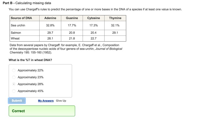
You can use Chargaff’s rules to predict the percentage of one or more bases in the DNA of a species if at least one value is known.
| Source of DNA |
Adenine |
Guanine |
Cytosine |
Thymine |
| Sea urchin |
32.8% |
17.7% |
17.3% |
32.1% |
| Salmon |
29.7 |
20.8 |
20.4 |
29.1 |
| Wheat |
28.1 |
21.8 |
22.7 |
|
|
Data from several papers by Chargaff: for example, E. Chargaff et al., Composition
of the desoxypentose nucleic acids of four genera of sea-urchin, Journal of Biological
Chemistry 195: 155-160 (1952). |
What is the %T in wheat DNA?
Part C
Use Chargaff’s rules to predict the missing values for E. coli, human, and ox DNA. Round your answers to the nearest whole number.
Part D – Evaluating a hypothesis
If Chargaff’s equivalence rule is valid, then hypothetically we could extrapolate this to the combined genomes of all species on Earth (as if there were one huge Earth genome). In other words, the total amount of A in every genome on Earth should equal the total amount of T in every genome on Earth. Likewise, the total amount of G in every genome on Earth should equal the total amount of C in every genome on Earth.
Calculate the average percentage for each base in your completed table. Do Chargaff’s equivalence rules still hold true when you consider those six species together?
Misconception Question 79
Part A
Meselson and Stahl cultured E. coli for several generations in a medium with a heavy isotope of nitrogen, 15N. They transferred the bacteria to a medium with a light isotope of nitrogen, 14 N. After two rounds of DNA replication, half the DNA molecules were light (both strands had 14N) and half were hybrids (15N-14N). What did the researchers conclude from these results?

Misconception Question 78
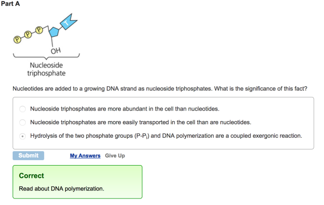
Part A

Nucleotides are added to a growing DNA strand as nucleoside triphosphates. What is the significance of this fact?
Misconception Question 80
Part A
Select the most accurate statement describing DNA replication complexes.

Misconception Question 77
Part A
During DNA replication, the leading strand is synthesized continuously, whereas the lagging strand is synthesized as Okazaki fragments. Why is this so?

Misconception Question 76
Part A
DNA is a self-replicating molecule. What accounts for this important property of DNA?

When cells divide or multiply, the DNA must be copied, or replicated. All known organisms replicate their DNA by a method known as semi-conservative replication, in which each of the two strands of the parent DNA becomes part of the daughter DNA.
Part A
Complete the following vocabulary exercise related to DNA replication.
Match the words in the left-hand column with the appropriate blank in the sentences in the right-hand column.

Part A
In E. coli replication the enzyme primase is used to attach a 5 to 10 base ribonucleotide strand complementary to the parental DNA strand. The RNA strand serves as a starting point for the DNA polymerase that replicates the DNA. If a mutation occurred in the primase gene, which of the following would you expect?
Part A
Hershey and Chase used a DNA-based virus for their work. What would the results have been if they had used an RNA virus?

Part A
The lagging strand is characterized by a series of short segments of DNA (Okazaki fragments) that will be joined together to form a finished lagging strand. The experiments that led to the discovery of Okazaki fragments gave evidence for which of the following ideas?

Part A
In his work with pneumonia-causing bacteria and mice, Griffith found that

Part A
What is the basis for the difference in how the leading and lagging strands of DNA molecules are synthesized?

Part A
In analyzing the number of different bases in a DNA sample, which result would be consistent with the base-pairing rules?

Part A
The elongation of the leading strand during DNA synthesis

Part A
In a nucleosome, the DNA is wrapped around

Part A
E. coli cells grown on 15N medium are transferred to 14N medium and allowed to grow for two more generations (two rounds of DNA replication). DNA extracted from these cells is centrifuged. What density distribution of DNA would you expect in this experiment?

Part A
A biochemist isolates, purifies, and combines in a test tube a variety of molecules needed for DNA replication. When she adds some DNA to the mixture, replication occurs, but each DNA molecule consists of a normal strand paired with numerous segments of DNA a few hundred nucleotides long. What has she probably left out of the mixture?

Part A
The spontaneous loss of amino groups from adenine in DNA results in hypoxanthine, an uncommon base, opposite thymine. What combination of proteins could repair such damage?

Activity: DNA Double Helix
Part A
In a DNA double helix an adenine of one strand always pairs with a(n) _____ of the complementary strand, and a guanine of one strand always pairs with a(n) _____ of the complementary strand.

Chapter 16 Pre-Test Question 3
Part A
Who conducted the X-ray diffraction studies that were key to the discovery of the structure of DNA?

Chapter 16 Pre-Test Question 2
Part A
In the Hershey and Chase experiment that helped confirm that DNA, not protein, was the hereditary material, what was the key finding?

Chapter 16 Pre-Test Question 1
Part A
Griffith’s experiments with S. pneumoniae were significant because they showed that traits could be transferred from one organism to another. What else did he find that was significant?

Activity: DNA Replication: A Review
Part A
Short segments of newly synthesized DNA are joined into a continuous strand by _____.

Part B
After DNA replication is completed, _____.

Part C
The first step in the replication of DNA is catalyzed by _____.

Part D
The action of helicase creates _____.

Part E
Why is the new DNA strand complementary to the 3′ to 5′ strands assembled in short segments?
Part F
The synthesis of a new strand begins with the synthesis of a(n) _____.
Part G
Which of these is responsible for catalyzing the formation of an RNA primer?


Part H
An old DNA strand is used as a _____ for the assembly of a new DNA strand.
Activity: DNA Replication: A Closer Look
Part A
In a DNA double helix an adenine of one strand always pairs with a(n) _____ of the complementary strand, and a guanine of one strand always pairs with a(n) _____ of the complementary strand.

Part B
After DNA replication is completed, _____.

Part C
The first step in the replication of DNA is catalyzed by _____.

Part D
The action of helicase creates _____.

Part E
Why is the new DNA strand complementary to the 3′ to 5′ strands assembled in short segments?

Part F
The synthesis of a new strand begins with the synthesis of a(n) _____.

Part G
An old DNA strand is used as a _____ for the assembly of a new DNA strand.

Activity: DNA Replication: An Overview
Part A
Short segments of newly synthesized DNA are joined into a continuous strand by _____.

Part B
After DNA replication is completed, _____.

Part C
The action of helicase creates _____.

Part D
Why is the new DNA strand complementary to the 3′ to 5′ strands assembled in short segments?

Part E
An old DNA strand is used as a _____ for the assembly of a new DNA strand.

Activity: DNA Synthesis
Part A
What catalyzes DNA synthesis?

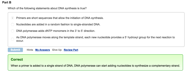
Part B
Which of the following statements about DNA synthesis is true?
Part C
Which part of a deoxynucleoside triphosphate (dNTP) molecule provides the energy for DNA synthesis?

Part D
Which of the following enzymes creates a primer for DNA polymerase?

Part E
Which of the following statements about Okazaki fragments in E. coli is true?

Part F
Which of the following enzymes is important for relieving the tension in a helix as it unwinds during DNA synthesis?

Part G
True or false? Single-stranded DNA molecules are said to be antiparallel when they are lined up next to each other but oriented in opposite directions.

DNA Replication: Mechanism and Proteins
DNA replication is the process by which DNA is copied. It is highly accurate in both bacteria and eukaryotes and requires a variety of DNA polymerases and other accessory proteins. In this tutorial you will learn how DNA is replicated and understand the roles of the proteins involved in the process.
Part A – The mechanism of DNA replication
The diagram below shows a double-stranded DNA molecule (parental DNA).
Drag the correct labels to the appropriate locations in the diagram to show the composition of the daughter DNA molecules after one and two cycles of DNA replication. In the labels, the original parental DNA is blue and the DNA synthesized during replication is red.
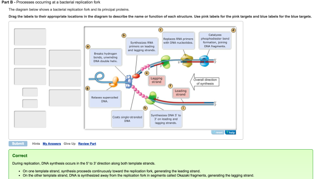
Part B – Processes occurring at a bacterial replication fork
The diagram below shows a bacterial replication fork and its principal proteins.
Drag the labels to their appropriate locations in the diagram to describe the name or function of each structure. Use pink labels for the pink targets and blue labels for the blue targets.
Chapter 16 Pre-Test Question 9
Part A
What are chromosomes made of?
Chapter 16 Pre-Test Question 10
Part A
Which of the following is true of DNA during interphase?


Wow you are a legend
LikeLiked by 1 person
Die ist ja total süß. Die nähe ich ganz bestimmt irgendwann mal nach!
LikeLike
Thank you!!!!!!!!!!!!!!!!!!!!!!!!!!!!!!
LikeLiked by 1 person
Thank you so much!
LikeLike
wow thanks so much I was struggling
LikeLike
Thank you so much for making this!!
LikeLike
AMAZING THANK Y
LikeLike
thanks for such a good definition can you please shed some light here i often confuse direct response advertising and direct response marketing?
van cleef and arpels turquoise alhambra necklace fake http://www.classicaljewelry.net/
LikeLike
hOur company provides healthcare products. Visit our health contributing site in case you want to feel healthier. Our company provides supreme quality health and related products. Take a look at our health contributing portal in case you want to look healthier. Our company provides a wide variety of healthcare products. Take a look at our health contributing website in case you want to look healthier. Our company offers herbal healthcare products. Look at our health contributing portal in case you want to look healthier. Our company provides a wide variety of non prescription products. Visit our health portal in case you want to look better with a help of generic supplements. Our company provides a wide variety of health products. Look at our health contributing website in case you want to look healthier.
Our company offers safe health and related products. Look at our health contributing portal in case you want to look healthier. Our company offers a wide variety of health and related products. Take a look at our health contributing site in case you want to look better. Our company offers herbal general health products. Look at our health contributing portal in case you want to look healthier. Our company offers a wide variety of non prescription products. Visit our health portal in case you want to look healthier with a help of generic supplements. Our company provides a wide variety of non prescription drugs. Visit our health portal in case you want to strengthen your health with a help general health products.
LikeLike
you are literally the best
LikeLike
THANK YOU THANK YOU THANK YOU!!!!!! I love biology but my teacher is making me fall out of love.
LikeLike
Pingback: Bioflix Activity: Mitosis- The Cell Cycle Part A – The Cell Cycle Drag The Pink Labels… - [100% Official Pages]